How to Use a Microscope
The eyepiece on a microscope magnifies at 10x, so when used together, the 4x lens magnifies an item 40x, the 10x magnifies 100x, and the 40x magnifies 400x. (note: for typical student microscope - other microscopes will vary)
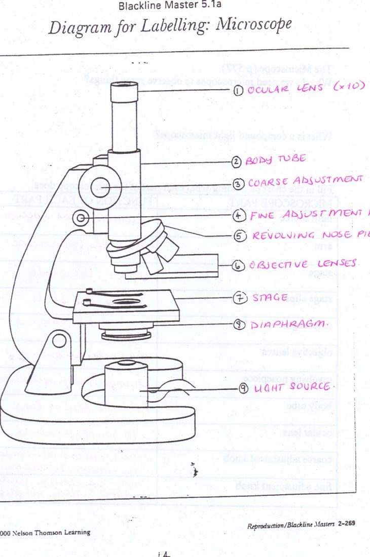
Monday September 25 Parts of a Compound Light Microscope
Download the Label the Parts of the Microscope PDF printable version here. Download the Label the Parts of the Microscope: Answers PDF printable version here. Microscope World explains the parts of the microscope, including a printable worksheet for schools and home.

Light Microscope Definition, Principle, Types, Parts, Labeled Diagram
Brian J. Ford Research biologist, Cambridgeshire, England, and fellow of Cardiff University, Wales. Author of Using the Digital Microscope and many books explaining and popularizing science. Brian J. Ford, Robert R. Shannon Emeritus Professor of Optical Sciences, University of Arizona, Tucson.
SCB 115 Lab 2 Microscope and pH, Acids, Bases, and Buffers Natural
Use this interactive to identify and label the main parts of a microscope. Drag and drop the text labels onto the microscope diagram.

Diagrams of a Microscope 101 Diagrams
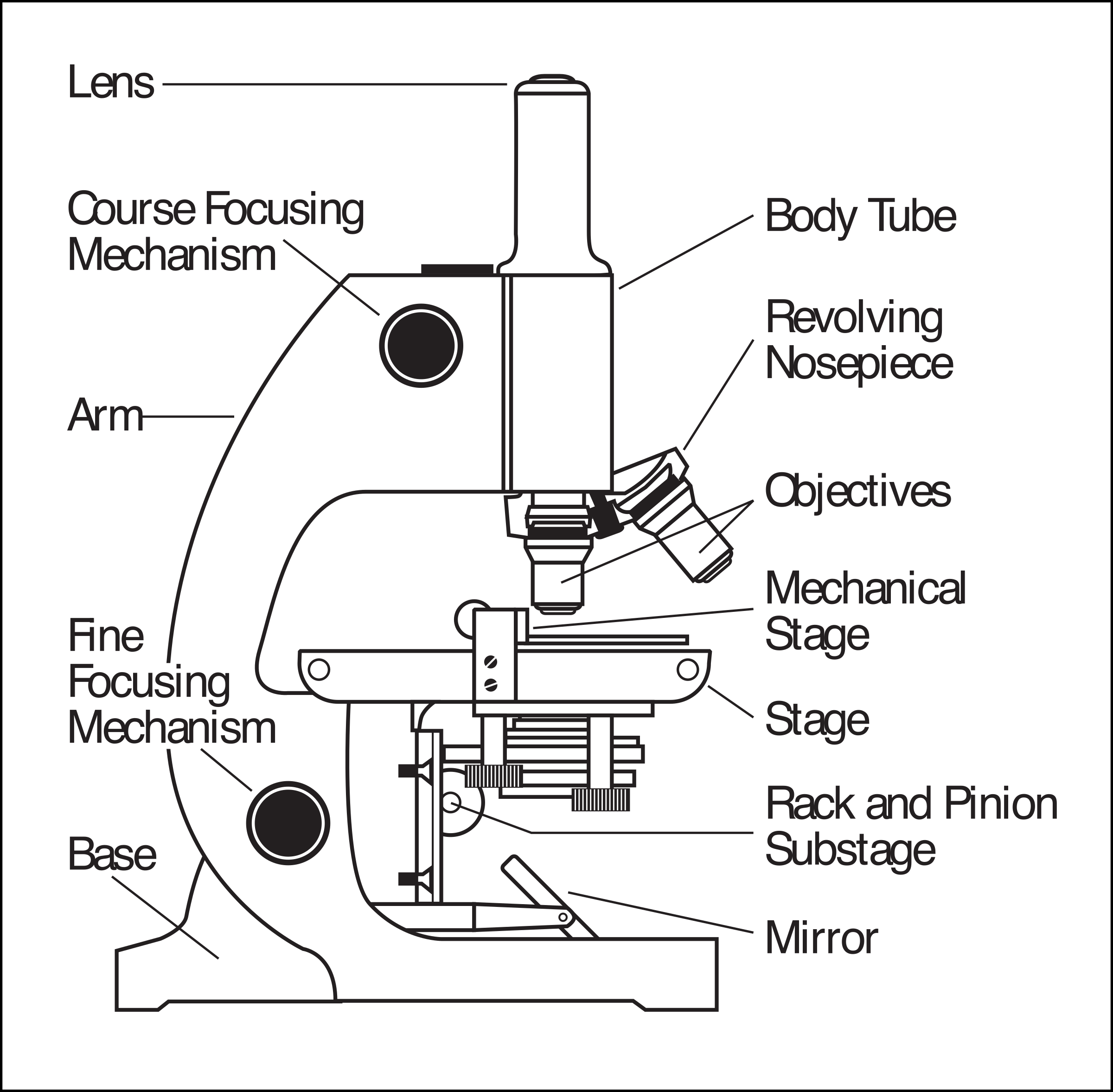
Microscope Parts Labeled Diagram The principle of the microscope gives you an exact reason to use it. It works on the three principles. Magnification Resolving Power Numerical Aperture. Parts of a Microscope

The Compound Microscope Diagram Microscopic Diagram Microscope Parts
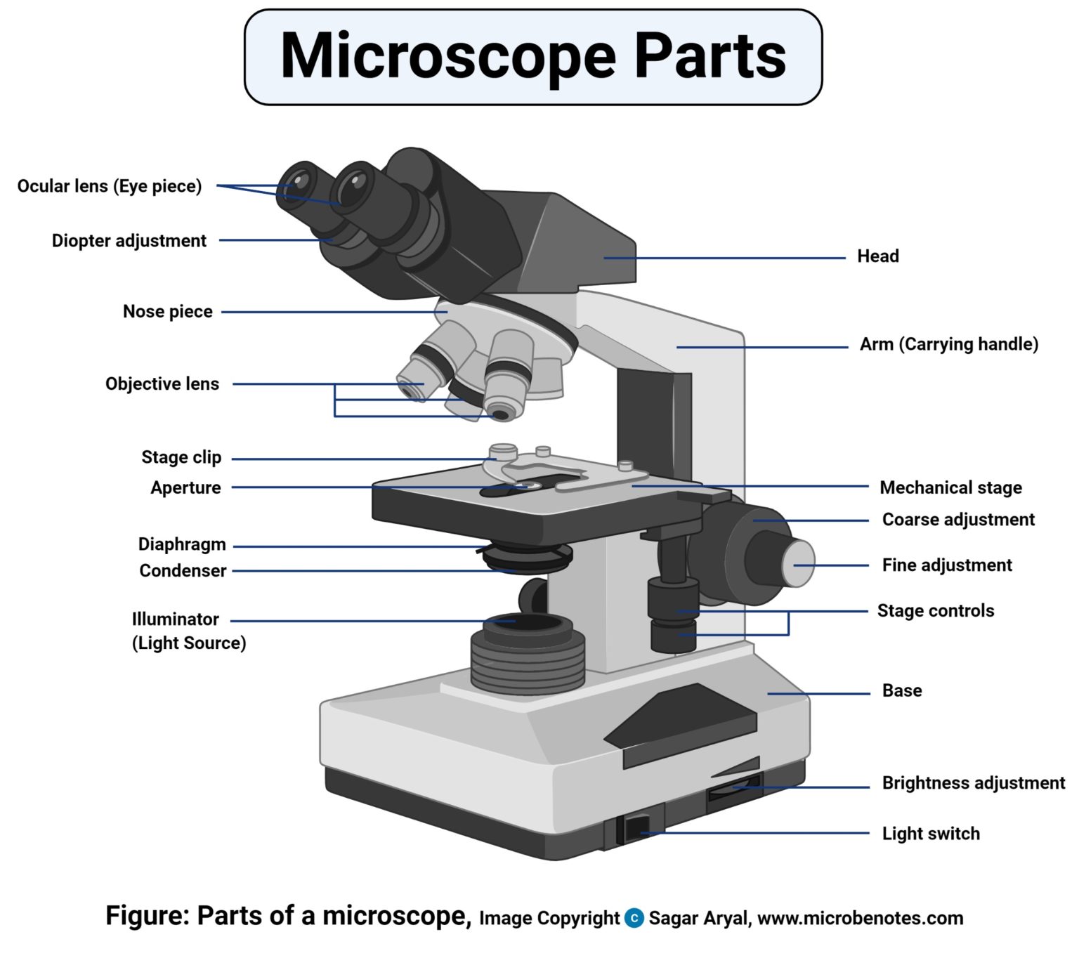
The optical microscope often referred to as the light microscope, is a type of microscope that uses visible light and a system of lenses to magnify images of small subjects. There are two basic types of optical microscopes: Simple microscopes. Compound microscopes. The term "compound" in compound microscopes refers to the microscope having.

Microscope labeled diagram
Labeled diagram Exploring Microscope Functions: Magnification is the technique of magnifying the picture of a specimen to make small structures apparent to the human eye. Magnification is often stated as a numerical value that indicates how many times bigger the picture is relative to the real size of the specimen.
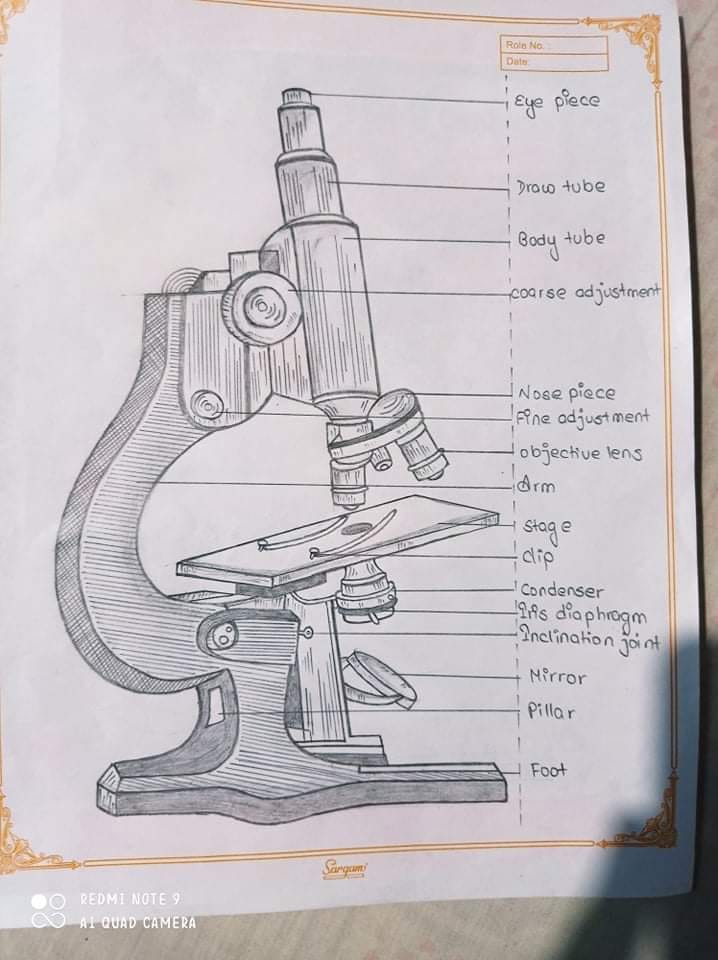
Aggregate 80+ microscope drawing with label nhadathoangha.vn
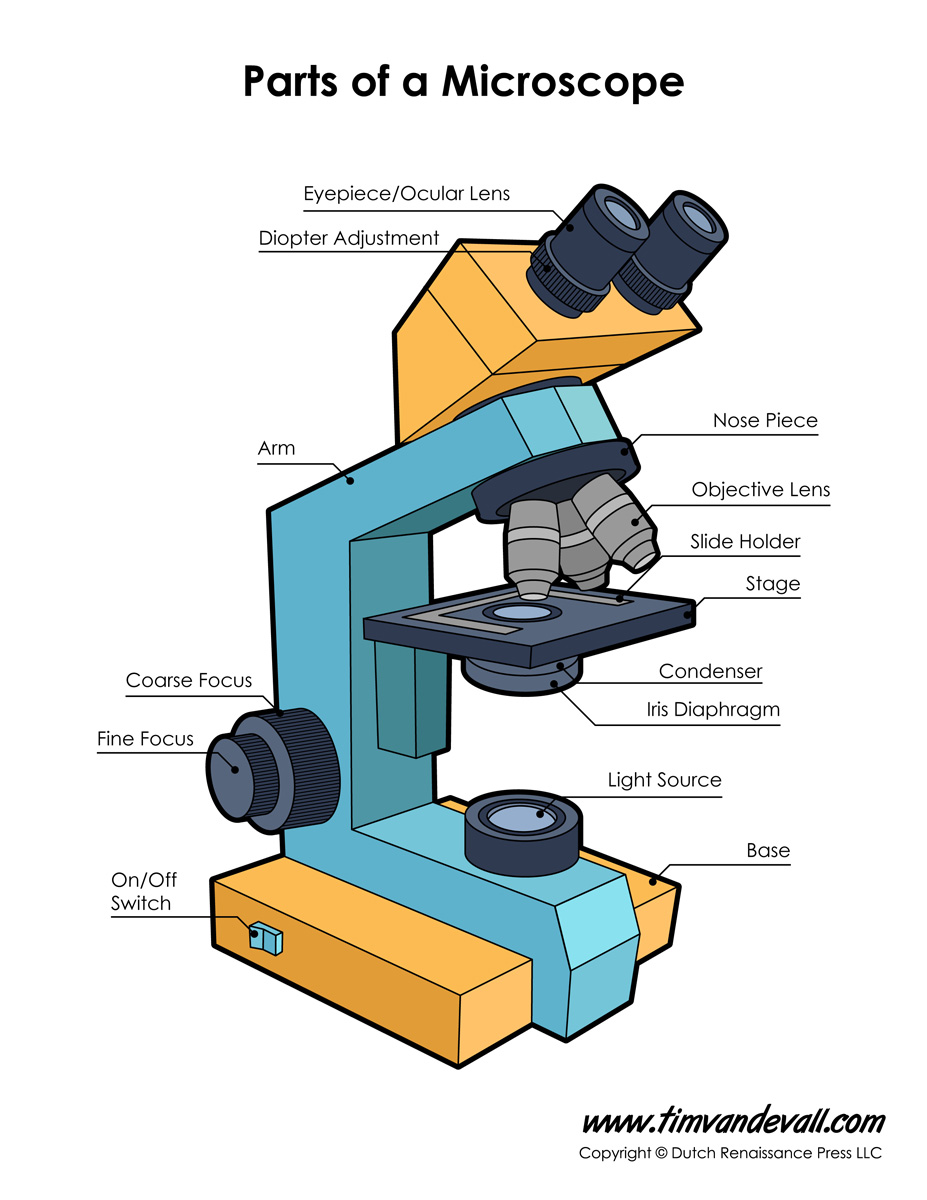
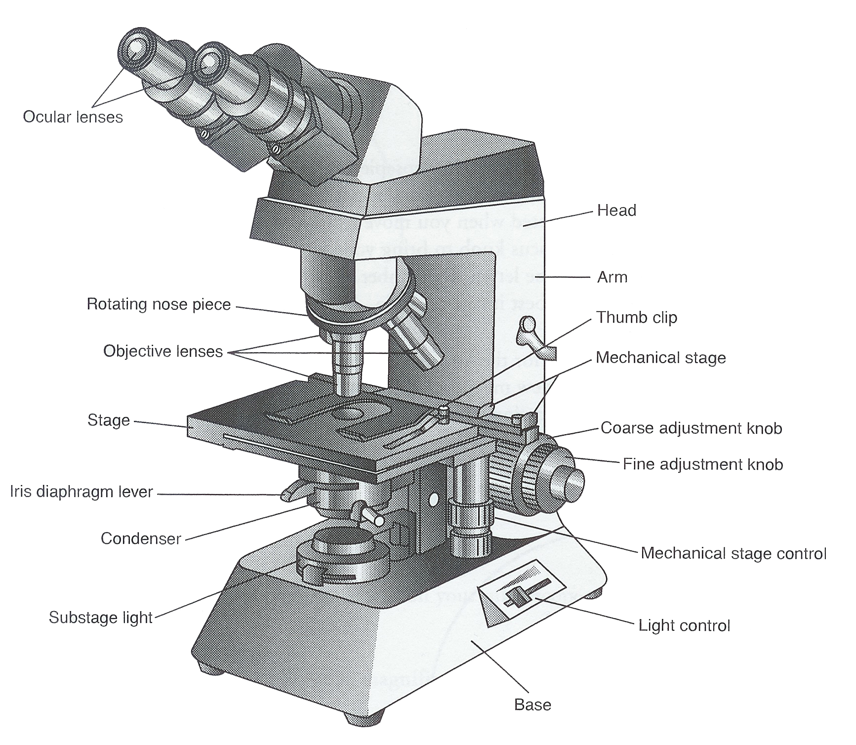
Labeled diagram of a compound microscope Major structural parts of a compound microscope Optical components of a compound microscope Eyepiece Eyepiece tube Objective lenses Nosepiece Specimen stage Coarse and fine focus knobs Rack stop Illuminator Condenser Abbe condenser Iris Diaphragm Condenser Focus Knob Summary An overview of microscopes
Microscope Diagram to Print 101 Diagrams
Introduction If you meet some cell biologists and get them talking about what they enjoy most in their work, you may find it comes down to one thing: secretly, they're all microscope freaks.
Labeled Microscope Diagram Ks3 Micropedia Gambaran
With Labeled Diagram and Functions How does a Compound Microscope Work? Before exploring microscope parts and functions, you should probably understand that the compound light microscope is more complicated than just a microscope with more than one lens.
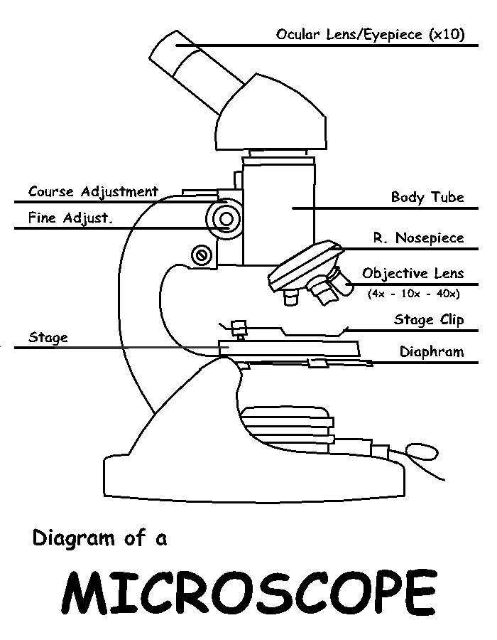
Diagram of a Microscope by ScienceDoodles on DeviantArt
The working principle of a simple microscope is that when a lens is held close to the eye, a virtual, magnified and erect image of a specimen is formed at the least possible distance from which a human eye can discern objects clearly. Magnification formula The magnification power of a simple microscope is expressed as: M = 1 + D/F Where

Label the Microscope Diagram Download Scientific Diagram
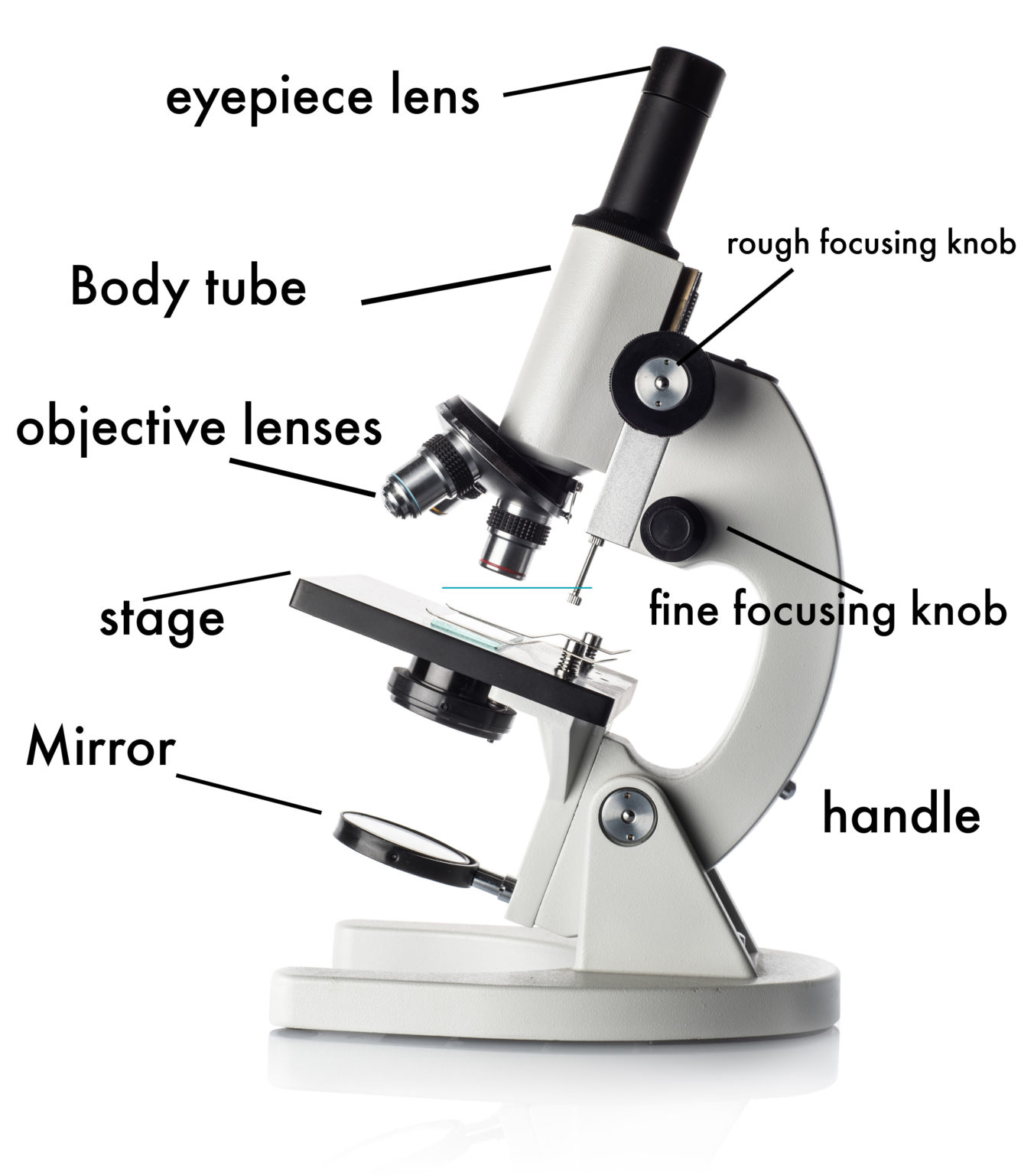
Parts of the Microscope with Labeling (also Free Printouts) A microscope is one of the invaluable tools in the laboratory setting. It is used to observe things that cannot be seen by the naked eye. Table of Contents 1. Eyepiece 2. Body tube/Head 3. Turret/Nose piece 4. Objective lenses 5. Knobs (fine and coarse) 6. Stage and stage clips 7. Aperture

Diagrams of Microscope 101 Diagrams
The most important parts of a microscope are the lenses, head, base, and arms. The lenses are in two locations: at the top inside the eyepiece and in the middle through the rotating objective.

36 Label Parts Of The Microscope Labels 2021
Parts of the Microscope (Labeled Diagrams) By Editorial Board December 14, 2022 The microscope is one of the must-have laboratory tools because of its ability to observe minute objects, usually living organisms that cannot be seen by the naked eyes. It is categorized into two: simple and compound microscopes.

Microscope Diagram to Print 101 Diagrams
They are: Light Microscopes: These use light rays to illuminate objects. e.g. Dissection microscopes and compound microscopes. Electron Microscopes: These illuminate objects with a beam of highly charged electrons. e.g. Transmission electron microscope (TEM) and scanning electron microscope (SEM).
Clipart microscope parts labeled WikiClipArt
Fluorescence microscopes: These use fluorescent dyes to highlight specific structures or molecules in a sample and are commonly used in biological research. X-ray microscopes: These use X-rays to produce images of the internal structure of samples and are often used to study materials and biological specimens.